An Atomic Force Microscope (AFM) is a very-high-resolution type of scanning probe microscopy (SPM) that allows for imaging and analysis of material surfaces at the nanoscale, down to fractions of a nanometer (atomic resolution). Unlike optical or electron microscopes, AFM "feels" the surface with a tiny probe, similar to how a finger explores textures, but at an incredibly small scale. How it Works: The fundamental principle behind AFM involves a sharp tip attached to a flexible cantilever, acting like a spring. Here's a breakdown of the key components and their function:
Cantilever and Tip: The heart of the AFM is a microfabricated cantilever with an extremely sharp tip (often silicon or silicon nitride) at its free end.
Sample Stage: The sample to be analyzed is mounted on a stage that can be precisely moved in three dimensions (x, y, and z) using piezoelectric actuators.
Laser and Photodetector: A laser beam is focused on the backside of the cantilever and reflects onto a position-sensitive photodetector (PSPD).
Feedback Loop and Electronics: The photodetector measures the cantilever's deflection (bending) or oscillation changes.
Operating Modes: AFM can operate in several modes, each suited for different sample types and measurements:
Contact Mode: The tip is in continuous contact with the sample surface.
Non-Contact Mode: The cantilever oscillates just above the sample surface, typically in the attractive force regime.
Tapping Mode (Intermittent Contact Mode): The cantilever oscillates at or near its resonant frequency, and the tip intermittently "taps" the sample surface at the lowest point of its oscillation.
Beyond Topography: While primarily known for its high-resolution topographical imaging, AFM is incredibly versatile and can be used to measure various local physical properties of materials by sensing different tip-sample interactions.
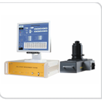 Hall Effect Meserment System
 Four-Point Probe
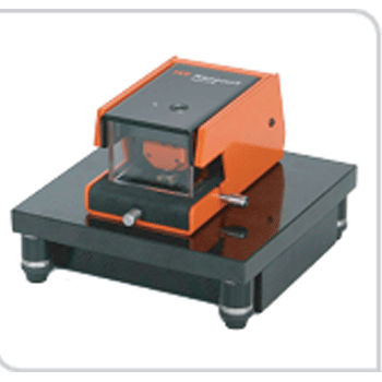
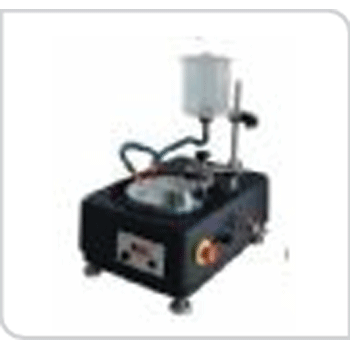 Polishing & Grainding Machine
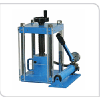 Lab Press With Hydraulic Pump
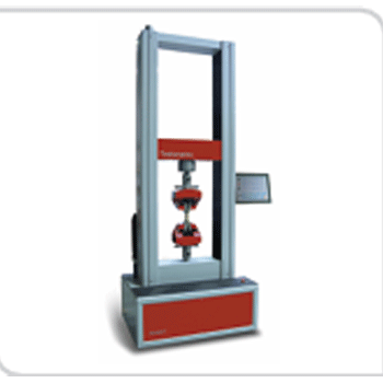 Universal Tensile Tester (UTM)